XB-IMG-212296
Xenbase Image ID: 212296
|
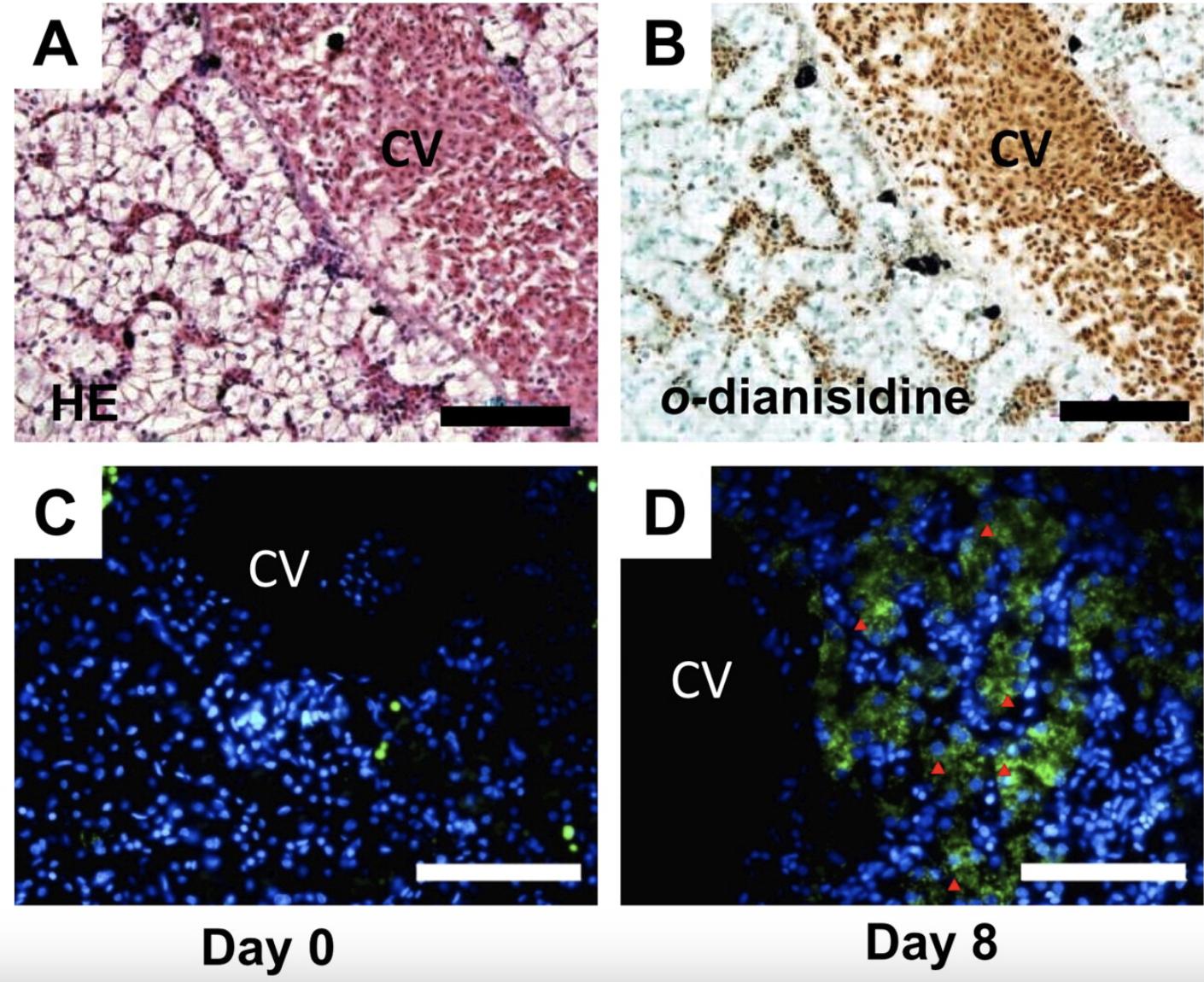
Fig. 1. Erythropoietin receptor (EPOR) expression in the adult liver of Xenopus laevis. Hematoxylin and eosin staining (A) and o-dianisidine staining (B) of paraffin-embedded sagittal tissue sections (4 m thickness) fixed in Bouin's solution. CV indicates the central vein. Immunohistochemical staining of normal (Day 0, C) and phenylhydrazine (PHZ)-anemic (Day 8, D) transverse liver sections (10 m thickness) with xlEPOR polyclonal antibody. xlEPOR-positive cells reside in the hepatic sinusoid of anemic liver. Arrowheads indicate xlEPOR-positive cells, which lie adjacent to the central vein. Nuclei are stained with DAPI (blue). Scale bars represent 100 m. Image published in: Nogawa-Kosaka N et al. (2011) Copyright © 2011. Image reproduced with permission of the Publisher.
Image source: Published Larger Image Printer Friendly View |